Medical Summary
When investigating what causes low testosterone in young males, clinical evaluation must distinguish between primary testicular failure, secondary central defects, and functional suppression. In men under thirty, a decline in androgen production is never considered a normal physiological consequence of ageing. Instead, it signals an underlying pathology or significant environmental stressor that demands thorough diagnostic workup. Primary hypogonadism in this demographic frequently stems from congenital conditions such as Klinefelter syndrome, anatomical issues like cryptorchidism, or direct testicular trauma. Conversely, secondary hypogonadism points towards hypothalamic-pituitary axis dysfunction, which may be triggered by prolactinomas, traumatic brain injury, or congenital gonadotropin-releasing hormone (GnRH) deficiencies.
MYTRT treatment eligibility
MYTRT treats men only when they have compatible symptoms and meet both biochemical limits: total testosterone of 15 nmol/L or below, and free testosterone of 0.35 nmol/L or below.
In clinic we see an increasing prevalence of functional hypogonadism driven by lifestyle and iatrogenic factors. The widespread use of anabolic-androgenic steroids (AAS) and selective androgen receptor modulators (SARMs) has become a leading cause of profound, sometimes irreversible, axis suppression in young men. Furthermore, metabolic syndrome, chronic opioid therapy, and severe psychological or physical stress can significantly blunt luteinising hormone (LH) pulsatility. Proper management requires a comprehensive biochemical assessment, including early morning total testosterone, free testosterone, LH, follicle-stimulating hormone (FSH), prolactin, and sex hormone-binding globulin (SHBG), to pinpoint the exact aetiology before any intervention is considered[1].
Understanding Hypogonadism in Young Men
The endocrine system relies on a delicate feedback loop involving the hypothalamus, the pituitary gland, and the testes. The hypothalamus releases GnRH, which signals the pituitary gland to secrete LH and FSH. These hormones then travel through the bloodstream to the testes, stimulating Leydig cells to produce testosterone and Sertoli cells to facilitate spermatogenesis. When this system breaks down in a young man, it is broadly categorised into three distinct types of hypogonadism: primary, secondary, and functional. Each of these categories requires a unique diagnostic approach and a tailored management strategy to ensure long-term health and wellbeing.
Primary hypogonadism occurs when the testes themselves fail to respond to adequate stimulation from the brain. The pituitary gland detects the low circulating androgens and attempts to compensate by increasing LH and FSH output, resulting in hypergonadotropic hypogonadism. Secondary hypogonadism, on the other hand, originates in the brain. The testes are perfectly capable of producing hormones, but they do not receive the necessary signals due to hypothalamic or pituitary dysfunction, leading to hypogonadotropic hypogonadism. Functional hypogonadism is a subset of secondary failure where the suppression is potentially reversible, often driven by external factors such as medication, extreme weight fluctuations, or severe systemic illness[2].
Identifying the precise mechanism is paramount because it dictates the clinical approach. While some men may require lifelong hormone replacement, others might restore their natural production by addressing the root cause. If you are experiencing signs of androgen deficiency, you can read more about low testosterone symptoms to understand how these biochemical changes manifest physically and psychologically. The symptoms can range from subtle changes in mood and energy to profound alterations in body composition and sexual function, making early detection and intervention critical.
Primary Hypogonadism: Testicular Causes
Primary testicular failure in young men is often linked to genetic anomalies, developmental issues, or direct physical damage to the gonads. In these cases, the Leydig cells are compromised, leading to insufficient androgen synthesis despite high levels of stimulating hormones from the pituitary gland. This hypergonadotropic state is a clear indicator that the problem lies within the testes themselves, necessitating a focused investigation into potential structural or genetic abnormalities.
1. Klinefelter Syndrome
Klinefelter syndrome is the most common congenital abnormality causing primary hypogonadism, affecting approximately one in every 600 male births. It occurs when a male is born with an extra X chromosome, resulting in a 47,XXY karyotype instead of the typical 46,XY. This genetic variation disrupts normal testicular development, leading to small, firm testes and progressive fibrosis of the seminiferous tubules and Leydig cells. The condition is often underdiagnosed, with many men only discovering they have it when investigating fertility issues or persistent symptoms of androgen deficiency in adulthood.
Young men with Klinefelter syndrome often present with delayed or incomplete puberty, gynaecomastia (enlarged breast tissue), reduced facial and body hair, and disproportionately long limbs. While some individuals may produce adequate androgens during early puberty, levels typically decline rapidly by their late teens or early twenties. Diagnosis is confirmed through karyotype testing, and early intervention is crucial to preserve bone density, support normal physical development, and mitigate the long-term metabolic risks associated with chronic androgen deficiency[1].
2. Undescended Testes (Cryptorchidism)
During foetal development, the testes form within the abdomen and normally descend into the scrotum shortly before birth. Cryptorchidism occurs when one or both testes fail to complete this descent. The scrotum provides a cooler environment essential for optimal testicular function; prolonged exposure to the higher core body temperature within the abdomen can cause irreversible damage to both the germ cells and the Leydig cells. This temperature sensitivity is a critical factor in maintaining both fertility and adequate hormone production throughout a man’s life.
Although surgical correction (orchidopexy) is usually performed in early childhood to bring the testes into the scrotum, individuals with a history of cryptorchidism remain at a higher risk of developing primary hypogonadism later in life. The severity of the impairment often correlates with the duration the testes remained undescended and whether the condition was bilateral. Regular monitoring of hormone profiles is recommended for young men with this medical history, as the decline in function can be gradual and insidious.
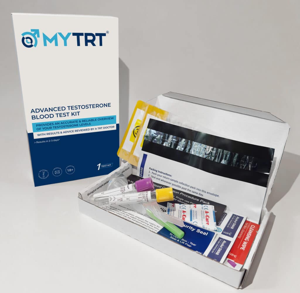
Advanced Testosterone Blood Test
Book a venous draw at a clinic £99.49
Book a venous draw at home with a nurse £109.49
 General health check
3. Testicular Injury or Torsion
The anatomical position of the testes makes them vulnerable to blunt force trauma, which can occur during contact sports, road traffic accidents, or occupational hazards. Severe trauma can rupture the testicle or compromise its blood supply, leading to tissue necrosis and subsequent failure of hormone production. Even seemingly minor injuries can sometimes result in long-term functional impairment if they cause significant inflammation or scarring within the delicate testicular structures.
Testicular torsion is a medical emergency where the spermatic cord twists, cutting off the arterial blood supply to the testicle. If surgical untwisting is not performed within a narrow window of a few hours, the testicle may suffer irreversible ischaemic damage and require removal (orchidectomy). While a single healthy testicle can often compensate and produce sufficient androgens, bilateral injury or the loss of a testicle in a man whose remaining testicle is already compromised will inevitably lead to a significant drop in circulating hormones[3].
4. Chemotherapy and Radiation Therapy
Advances in oncology have significantly improved survival rates for childhood and adolescent cancers, such as leukaemia, lymphoma, and testicular cancer. However, the treatments used to eradicate malignant cells can have profound gonadotoxic effects. Chemotherapeutic agents, particularly alkylating agents, are notorious for damaging the rapidly dividing cells within the testes. The extent of the damage is often dose-dependent and varies according to the specific drug regimen employed during treatment.
Radiation therapy directed at the pelvic region, lower abdomen, or total body irradiation can also cause extensive scarring and fibrosis of testicular tissue. The Leydig cells are generally more resilient to radiation than the germ cells responsible for sperm production, but high cumulative doses will eventually impair androgen synthesis. Young male cancer survivors require long-term endocrine follow-up to detect and manage late-onset hypogonadism, ensuring that their quality of life is maintained long after their cancer treatment has concluded.
5. Mumps Orchitis
Mumps is a highly contagious viral infection that primarily affects the salivary glands. However, if a male contracts mumps during or after puberty, the virus can spread to the testes, causing a painful inflammatory condition known as mumps orchitis. This complication occurs in up to 30% of post-pubertal males infected with the mumps virus, highlighting the importance of vaccination in preventing severe long-term reproductive and endocrine consequences.
The intense inflammation and subsequent swelling within the rigid tunica albuginea (the fibrous covering of the testis) can lead to pressure-induced ischaemia and atrophy of the testicular tissue. While bilateral mumps orchitis is less common than unilateral involvement, it carries a substantial risk of permanent primary hypogonadism and infertility. Widespread vaccination programmes have drastically reduced the incidence of mumps, but outbreaks still occur among unvaccinated populations, making this a relevant consideration in clinical practice.
Secondary Hypogonadism: Pituitary and Hypothalamic Causes
Secondary hypogonadism in young men indicates a failure at the level of the brain. The testes are structurally sound but remain dormant because they are not receiving the necessary gonadotropin signals (LH and FSH) from the pituitary gland. This central failure can be caused by structural lesions, genetic mutations, or severe trauma, and it requires a distinct diagnostic approach focused on neuroendocrine function rather than testicular anatomy.
6. Pituitary Disorders and Tumours
The pituitary gland, often referred to as the master gland, sits at the base of the brain and regulates multiple endocrine axes. Any structural abnormality, inflammatory condition, or space-occupying lesion within or adjacent to the pituitary can disrupt its function. Pituitary adenomas are benign tumours that can compress the surrounding healthy glandular tissue, impairing the secretion of LH and FSH. These tumours can grow silently for years before causing noticeable symptoms, making them a critical consideration in the differential diagnosis of secondary hypogonadism.
Other conditions affecting the pituitary include haemochromatosis (iron overload), sarcoidosis (inflammatory granulomas), and histiocytosis. In haemochromatosis, excess iron deposits accumulate in the pituitary gland, causing oxidative damage and cellular death. Young men presenting with secondary hypogonadism alongside symptoms of other pituitary hormone deficiencies (such as hypothyroidism or adrenal insufficiency) require urgent neuroimaging and comprehensive endocrine evaluation to rule out these potentially life-threatening conditions[4].
7. Prolactinoma
A prolactinoma is a specific type of functioning pituitary adenoma that secretes excessive amounts of prolactin. Prolactin is a hormone primarily associated with lactation in women, but it is also present in men. Hyperprolactinaemia (elevated prolactin levels) exerts a direct inhibitory effect on the hypothalamus, suppressing the pulsatile release of GnRH. This suppression is a potent mechanism for shutting down the entire reproductive axis, leading to profound hypogonadism.
Without GnRH, the pituitary ceases to produce LH and FSH, leading to a profound drop in androgen levels. Young men with a prolactinoma may present with visual field defects (due to the tumour pressing on the optic chiasm), headaches, gynaecomastia, and galactorrhoea (milky nipple discharge). Treatment typically involves dopamine agonist medications, which shrink the tumour and normalise prolactin levels, often restoring the hypothalamic-pituitary-gonadal (HPG) axis without the need for exogenous hormones.
8. Congenital GnRH Deficiency (Kallmann Syndrome)
Congenital hypogonadotropic hypogonadism (CHH) encompasses a group of rare genetic disorders characterised by the failure of the hypothalamus to produce or release GnRH. The most well-known form is Kallmann syndrome, which is distinguished by the additional symptom of anosmia or hyposmia (a complete or partial lack of the sense of smell). This unique combination of symptoms is a hallmark of the condition and aids in its clinical identification.
During embryonic development, the neurons responsible for secreting GnRH migrate from the olfactory placode to the hypothalamus. In Kallmann syndrome, a genetic mutation disrupts this migration pathway. Consequently, the individual is born without functional GnRH neurons. Young men with this condition typically fail to undergo spontaneous puberty and retain pre-pubertal testicular volume. Diagnosis is usually made in late adolescence when the absence of pubertal development becomes apparent, prompting a thorough endocrine and genetic investigation.
9. Head Injury and Traumatic Brain Injury (TBI)
The pituitary stalk, which connects the hypothalamus to the pituitary gland, is a delicate structure vulnerable to shearing forces during a traumatic brain injury. Road traffic collisions, severe falls, or repeated concussive impacts in contact sports (such as rugby or boxing) can disrupt the vascular supply or sever the neural connections between the brain and the pituitary. The increasing awareness of sports-related concussions has highlighted the importance of monitoring endocrine function in athletes who have sustained significant head trauma.
Post-traumatic hypopituitarism can manifest months or even years after the initial injury. The gonadotropic axis is often the most sensitive to trauma, making secondary hypogonadism a common complication of severe TBI. Young men with a history of significant head trauma who subsequently develop symptoms of lethargy, mood disturbances, or sexual dysfunction should be screened for pituitary dysfunction, as early intervention can significantly improve their recovery and quality of life.
Functional and Lifestyle-Driven Causes
Functional hypogonadism occurs when the HPG axis is suppressed by external, often modifiable, factors. In men under thirty, lifestyle choices, medications, and metabolic health play a massive role in determining endocrine function. Addressing these underlying issues can sometimes reverse the suppression and restore natural hormone production, which is why understanding how to increase testosterone naturally is a critical first step for many patients before considering pharmacological interventions.
10. Obesity and Metabolic Syndrome
The global rise in obesity among young adults has led to a parallel increase in functional hypogonadism. Adipose tissue (body fat) is not merely an inert energy store; it is an active endocrine organ that expresses the enzyme aromatase. Aromatase converts circulating androgens into ooestradiol (a form of oestrogen). Elevated oestrogen levels send a negative feedback signal to the brain, suppressing the release of GnRH, LH, and FSH. This hormonal imbalance can have far-reaching effects on a young man’s physical and mental health.
Furthermore, obesity is closely linked to insulin resistance and systemic inflammation, both of which independently impair Leydig cell function and reduce SHBG levels. This creates a vicious cycle: low androgens promote fat accumulation, and excess fat further suppresses androgen production. Weight loss through dietary modification and exercise is the primary intervention for obesity-induced hypogonadism, and significant improvements in hormone levels can often be achieved with sustained lifestyle changes[5].
11. Anabolic Steroid and SARM Use
The illicit use of anabolic-androgenic steroids (AAS) and selective androgen receptor modulators (SARMs) for physique enhancement is an epidemic among young men. When exogenous androgens are introduced into the body, the hypothalamus and pituitary gland detect the artificially high hormone levels and completely shut down the production of LH and FSH. This leads to profound testicular atrophy and a cessation of endogenous hormone synthesis, a condition that can be devastating for a young man’s long-term health.
Patients often ask us how long it takes to recover after stopping these compounds. The reality is that anabolic steroid-induced hypogonadism (ASIH) can persist for months or even years after cessation. In some cases, particularly with prolonged use of high doses, the HPG axis may never fully recover, leaving the individual dependent on medical intervention. Post-cycle therapy (PCT) protocols are often employed illicitly, but they do not guarantee a return to baseline function, and professional medical guidance is essential for managing the complex endocrine fallout of AAS or SARM use[6].
12. Chronic Stress, Overtraining, and Poor Sleep
The modern lifestyle often involves high levels of psychological stress, inadequate sleep, and sometimes extreme physical exertion. Chronic stress elevates cortisol levels, which directly inhibits the HPG axis at multiple levels, reducing both GnRH pulsatility and Leydig cell responsiveness. Similarly, severe caloric restriction and overtraining syndrome (often seen in endurance athletes or combat sports competitors cutting weight) signal to the body that it is in a state of starvation, prompting it to shut down non-essential functions like reproduction in order to conserve energy.
Sleep architecture is equally critical. The majority of daily androgen production occurs during the first deep phase of rapid eye movement (REM) sleep. Chronic sleep deprivation, shift work, or undiagnosed obstructive sleep apnoea can severely truncate this production window, leading to clinically significant hormonal deficits in otherwise healthy young men. Prioritising sleep hygiene and stress management is a fundamental component of restoring endocrine balance and overall wellbeing.
Diagnostic Workflow: Identifying the Cause
When a young man presents with symptoms of androgen deficiency, a structured diagnostic approach is essential. The initial step involves a comprehensive blood panel drawn in the early morning (between 8:00 AM and 10:00 AM) when hormone levels peak. If the total or free levels are found to be low, the test must be repeated to confirm the result, as transient fluctuations are common and a single low reading is insufficient for a definitive diagnosis.
Once a deficiency is confirmed, the clinician will evaluate the gonadotropins (LH and FSH) to categorise the condition. The table below outlines the typical biochemical patterns used to differentiate the aetiology, providing a clear framework for clinical decision-making.
| Condition Type | Testosterone Level | LH and FSH Levels | Clinical Interpretation |
|---|---|---|---|
| Primary Hypogonadism | Low | High (Elevated) | Testicular failure; the brain is signalling strongly, but the testes cannot respond. |
| Secondary Hypogonadism | Low | Low or Inappropriately Normal | Central failure; the brain is failing to send the necessary signals to the testes. |
| Functional Hypogonadism | Low | Low or Inappropriately Normal | Suppression due to lifestyle, obesity, or medications; potentially reversible. |
| Compensated Hypogonadism | Normal (Low-Normal) | High (Elevated) | Early-stage testicular strain; the brain is working harder to maintain normal levels. |
Further investigations may include a prolactin test, an iron panel (to rule out haemochromatosis), karyotyping (if Klinefelter syndrome is suspected), or an MRI of the pituitary gland to check for adenomas. A comprehensive testosterone test is the foundation of this diagnostic workflow, providing the critical data needed to formulate a safe and effective management plan tailored to the individual’s specific needs. A thourough history including pubertal development essential in under 30s, and this represents the smallest group of men undergoing tesosterone therapy medically.
Testosterone Blood Test Kit

If this test showed you have low testosterone, the Advanced Testosterone Blood Test will assess your suitability for TRT.
When to Seek Medical Advice
Experiencing a decline in vitality, libido, or physical performance in your twenties or thirties is not something to ignore or attribute solely to stress. Because low androgens in this age group are highly indicative of an underlying medical issue, prompt investigation is vital. Delaying diagnosis can lead to long-term consequences, including irreversible bone density loss, metabolic deterioration, and prolonged psychological distress that can impact every aspect of a young man’s life.
If you have a history of testicular trauma, previous anabolic steroid use, or are experiencing persistent symptoms, you should consult a specialist. Depending on the precise cause identified during the diagnostic workup, your clinician will discuss the most appropriate path forward. For some, lifestyle modifications may suffice, while others may require formal low testosterone treatment to restore their health and quality of life. The key is to take proactive steps towards understanding your body and seeking professional guidance when necessary.
References
3. BSSM , Guidelines on adult testosterone deficiency
4. Endocrine Society , Testosterone Therapy in Men With Hypogonadism